Mouse model instabilities 25/08/15
Progress: Simulations with conservative parameters of delta t have still been yielding unstable results.
As proliferation parameter rho is slowed down, the tumours look more real on the visualisation tools, to an extent. They never grow to fill the whole brain, the ones that look solid even have an instability where they start shrinking and the concentration drops to zero, which implies an unstable data point somewhere in the tensor(?)
e.g.


The simulations are taking now 40+ minutes to run on my desktop PC and are becoming unrunnable apart from for time scales longer than about 23 days on my MacBook.
Data files are very large now due to the higher resolution so until I get stable solutions, I am just saving a record of the parameters I have varied with notes on how the solution behaved.
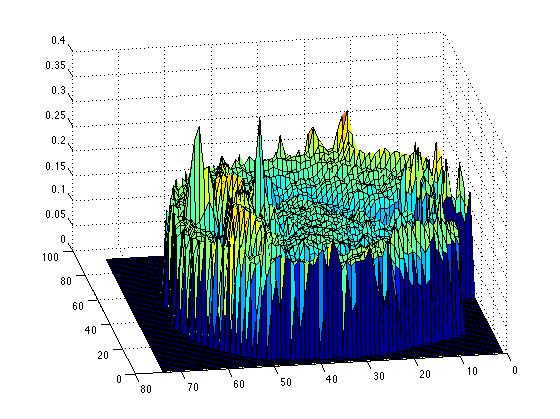
 Looking at the diffusion tensor variable Mean_Diff, surface plot of slice (:,:,101), I will try running simulations at around (40,40,101) which will be away from the two spikes, although they are only a factor of two or so larger than the surrounding points.
Data files are very large now due to the higher resolution so until I get stable solutions, I am just saving a record of the parameters I have varied with notes on how the solution behaved.